TISSUE, Class 9 notes
TISSUES
Tissue
can be defined as a group of cells with similar shape and function are termed
as tissues. They form a cellular organisational level, intermediate between the
cells and organ system. Organs are then created by combining the functional
groups of tissues.
Types
of Plant Tissues
The
classification of plant tissues are mainly based on the two important criteria:
- Based on the
different part of plants.
- Based on the
different types of cells.
Plant Tissues are
broadly categorised into three tissue systems. This classification is on
the basis of parts of the plants they are present.
- Epidermis
Tissues – cells formed from the outermost surface of the leaves.
- Vascular Tissues
– involved in transporting fluid and nutrients internally.
- Ground Tissue –
involved in producing nutrients by photosynthesis and preserve
nutrients.
Plant
tissue is divided into two types. This classification is on the basis of
the types of cells, they comprise.
- Meristematic
tissues.
- Permanent
tissues.
Meristematic Tissue
Meristematic
tissue is the plant tissue that has the ability to divide actively throughout
its life. They are the group of young cells, which consists of continually
dividing cells and helps in the increase of length and width of the plant.
Characteristics of Meristematic Tissue
1. The cells of these tissues are
commonly called meristems. The zone where these cells exist is known as
meristem.
2. Meristematic tissues contain living
cells with varied shapes
3. They possess a large nucleus devoid of
the vacuole.
4. The cells have no intercellular space.
5. The meristematic tissue has the
quality of self-renewal. Every time the cell divides, one cell remains
identical to the parent cell, and the others form specialized structures.
6. They have very small and few vacuoles.
7. The meristematic tissue is living and
thin-walled.
8. The protoplasm of the cells is very
dense.
9. The meristematic tissues heal the
wounds of an injured plant.
10. The cells of the meristematic tissue
are young and immature.
11. They do not store food.
12. They exhibit a very high metabolic
activity.
13. They possess a single, large and
prominent nucleus.
Functions
of Meristematic Tissue
- It is
responsible for the growth of the new organs.
- Involved in the
movement of water and nutrition within the plants.
- These tissues
are responsible for both primary and secondary growth of the plant.
- It is the
outermost tissue, functions by providing protection from mechanical injury.
- It gives rise to
epidermis layer, cortex, endodermis, ground tissue and vascular tissue.
There are different types of
meristematic tissues, which are classified on the basis of positions, functions,
plane of divisions, origin and development. The three main types of
meristematic tissues depending on the occurrence of the meristematic tissue on
the plant body are:
- Apical Meristem.
- Lateral
Meristem.
- Intercalary
Meristem.

Apical
Meristem
·
These
are present at the tips of the roots and shoots and helps in the increase of
the height of the plants.
·
Various
cell divisions facilitate the growth of the cells in the roots and shoots and
help in cellular enlargement.
·
Apical
meristem is divided into-promeristem zone, which contains actively dividing
cells, and the meristematic zone, which contains protoderm, procambium and
ground meristem.
Intercalary
Meristem
·
It
is located in the leaves and internodes at the intercalary position.
·
These
help to increase the length of the internode.
·
It
is found in grass, monocots and pines.
·
It
is a part of apical meristem and adds to the height of the plant.
Lateral
Meristem
·
It
is located in the stems and roots on the lateral side.
·
It
increases the thickness of the plant.
·
Vascular
cambium and cork cambium are the two lateral meristems.
·
These
divide preclinically or radially and give rise to secondary permanent tissues.
Permanent Tissues
A group of
cells which are similar in origin, structure and in function. They are involved
in complete growth and differentiation during the ineffective of meristematic
activity. There are two types of permanent tissues:
- Simple Permanent
Tissues.
- Complex
Permanent Tissues.
Functions
of Permanent Tissues
- In aquatic
plants, these tissues help in floating.
- Stores food in
the form of starch, proteins, oils and fats.
- They provide
hardness to fruits such as nuts, coconut, almond etc.
- These tissues
contain chloroplast which helps in carrying out photosynthesis.
- Permanent
Tissues are also involved in the Secretion, Transportation, and provides
mechanical support to the plants.
Types
of Permanent Tissue
Simple Permanent Tissue
These are
also known as homogenous tissues. They are made up of a single cell type,
usually with the same origin, structure, and function.
Simple
permanent tissue is further classified into three types:
Parenchyma
- The cells have
an oval or round shape.
- The cell wall is
made up of hemicellulose or cellulose.
- The cell is
thin-walled.
- The cells have
vacuoles and very small nucleus.
- It is found in
all parts of the plant.
- The protoplasm
is living and dense.
Collenchyma
- Cells are long
and thick-walled.
- The cell wall is
made up of cellulose and pectin.
- It is the only
tissue with the highest refractive index due to the presence of pectin.
- It is found in
the epidermis and the vascular bundle of dicot leaf.
- The amount of
chloroplast is less in the cells.
- The cells have
no intercellular spaces.
Sclerenchyma
- These are dead
tissues, very hard and rigid in texture.
- Cells are
thick-walled with various size and shapes.
- These provide
mechanical support and rigidity to the plant.

Complex permanent Tissue
The
complex tissues are made up of various types of cells carrying out distinct
functions and are of two types:
Xylem
- It transports
water and nutrients from the roots to the leaves of the plant.
- It provides
support to the plants.
- It is divided
into-tracheids, vessels, xylem fiber, and xylem parenchyma.

Phloem
- It translocates
the prepared organic food from the leaves to different parts of the plant.
- It is also known
as bast.
- It is composed
of sieve tubes, companion cells, phloem parenchyma, and phloem fibres.


Differences between Xylem and Phloem
|
Xylem |
Phloem |
|
Definition |
|
|
Xylem tissues are the
tubular-shaped structure, with the absence of cross walls. This tissue
resembles the shape of a star. |
Phloem tissues are tubular-shaped,
elongated, structures with the presence of walls with thin sieve tubes. |
|
Location |
|
|
It is located in the centre of the
vascular bundle. |
It is located on the outer side of
the vascular bundle. |
|
Fibres |
|
|
Xylem fibres are smaller. |
Phloem fibres are larger. |
|
Found In |
|
|
They are present in roots, stems
and leaves. |
They are present in stems and
leaves, which later transports and grow in roots, fruits and seeds. |
|
Movements |
|
|
These tissues move in a
Unidirectional. (only in one direction – upward direction) |
These tissues move in a
Bidirectional. (both ways – up and down) |
|
Comprises |
|
|
They live with hollow dead cells. |
They live with cytoplasm without
the nucleus. |
|
No of Tissues |
|
|
The total amount of xylem tissue
is more. |
The total amount of phloem tissue
is less. |
|
Features |
|
|
It consists of tracheids, vessel
elements, xylem parenchyma, xylem sclerenchyma and xylem fibres. |
It consists of four elements:
companion cells, sieve tubes, bast fibres, phloem fibres, intermediary cells
and the phloem parenchyma. |
|
Functions |
|
|
Transports soluble mineral
nutrients and water molecules from the roots to the aerial parts of the
plant. |
Transports food and other
nutrients including sugar and amino acids from leaves to storage organs and
growing parts of the plant. |
|
Vascular Bundles |
|
|
Forms vascular bundles with
phloem. |
Forms vascular bundles with xylem. |
|
Functions |
|
|
Provides mechanical strength to
the plant and helps in strengthening the stem. |
Translocates the synthesized
sugars by the photosynthetic areas of plants to storage organs like roots,
bulbs and tubers. |
|
Functions |
|
|
It is responsible for replacing
the total amount of lost water molecules through transpiration and
photosynthesis. |
It is responsible for transporting
proteins and mRNAs throughout the plant. |
Animal Tissue
A
group of cells similar in structure, function, and origin is called tissues. In animals, the structure of tissue depends on its function.
An animal body is made of four different types of tissues. They have been
classified based on the type of cell, function, and location in the body. They
include:
- Epithelial Tissue
- Muscle Tissue
- Connective Tissue
- Nerve Tissue
A) Epithelial Tissue
All
layers and organs in the body are lined by a group of tissues called epithelial
tissues which are commonly referred to as epithelium. They cover the surface of
all internal as well as external organs. Epithelial tissue is highly permeable.
Thus, it plays a significant role in the exchange of substances across the
cells and helps in maintaining the osmoregulation. Depending on the number of
layers of cells it is composed of, the epithelium has been divided into the
simple epithelium and compound epithelium. The main functions of epithelial
tissue are protection, secretion, absorption, and sensation.
Squamous
Epithelium
Squamous epithelium form an extremely thin and
flat layer of tissues. They are semi-permeable and thus, perfect for gaseous
exchange. They are present in the lining of oesophagus and mouth.
Cuboidal
Epithelium
As the name suggests, they are cuboidal in shape
and form the lining of salivary glands and kidney tubules. They provide
mechanical support. They also form glandular epithelium when they form glands.
Columnar
Epithelium
These tissues line the organs which help in
absorption and secretion, such as lining of intestines. They are made up of
elongated cells. When cilia is present on these cells, they form ciliated
columnar epithelium like those present in the respiratory tract.
Stratified
Squamous Epithelium
This kind of tissue is formed when multiple
layers of squamous epithelium are arranged in a pattern. Our skin is made up of
this kind of tissue.

B) Muscular Tissue
These tissues make up our muscles which are
responsible for almost all the movements that take place in the body.
Properties of Muscular Tissue
1. Contractibility– It is the ability of muscle cells to
shorten forcefully.
2. Extensibility– A muscle has the ability to be
stretched.
3. Elasticity– The muscles have the ability to
recoil back to its original length after being stretched.
4. Excitability– The muscle tissue responds to a
stimulus delivered from a motor neuron or hormone.
Types of Muscular Tissue
The
muscular tissue is of three types:
·
Skeletal
Muscle Tissue
·
Smooth
Muscle Tissue
·
Cardiac
Muscle Tissue
Skeletal Muscle Tissue

·
These muscles are attached to the
skeleton and help in its movement.
·
These
muscles are also known as striated muscles because of the presence of alternate
patterns of light and dark bands.
·
These
light and dark bands are sarcomeres which are highly organized structures of
actin, myosin, and proteins. These add to the contractility and extensibility
of the muscles.
·
Skeletal muscles are voluntary muscles composed
of muscle fibers.
·
40%
of our body mass comprises skeletal muscles.
·
Each
skeletal tissue contains myofibrils.
·
The
cells of these tissues are multinucleated.
·
These
are provided with blood vessels and many elongated mitochondria and glycogen
granules.
·
They
bring about the movement of the organs of the body.
·
They are long, cylindrical, unbranched with striations and
are multinucleated.
·
They are called skeletal because these tissues are mostly
attached to the bones.
Smooth
Muscle Tissue

·
These
are non-striated, involuntary muscles controlled by the Autonomous Nervous
System.
·
It
stimulates the contractility of the digestive, urinary, reproductive systems,
blood vessels, and airways.
·
The actin and myosin filaments are
very thin and arranged randomly, hence no striations.
·
The
cells are spindle-shaped with a single nucleus.
·
They are long, smooth, spindle shaped and uninucleate.
Cardiac Muscle Tissue
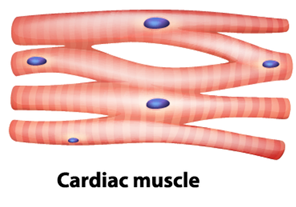
·
These
are found only in the heart.
·
These
are involuntary muscles and the heart pumps the blood through cardiac
contractions.
·
The
cells of the cardiac muscles known as the cardiomyocytes are striated.
·
They
are single-celled and uninucleated.
- The ends of the
cells are joined and the junctions are called intercalated discs. The
cells are attached to each other by desmosomes.
- Structurally
they may look quite similar to striated muscles but they are branched,
uninucleate and have intercalated discs.
- These
muscles are involuntary in nature and show rhythmic contractions and
relaxations.,
|
Cardiac muscle |
Striated muscle |
|
Cardiac muscles are multinucleate
and branched |
Striated muscles are multinucleate
and unbranched |
|
These are involuntary |
These are voluntary |
|
These are found in the heart |
Attached
to bones of the entire body |
|
Shorter in length |
Longer in length |
|
Controlled by Autonomic nervous system |
controlled by somatic nervous system |
|
Semi-spindle |
cylindrical |
|
Help in Pumping
blood |
Help in Movement of the body |
|
Gap junctions present |
Gap junctions absent |
|
Speed of contraction is fast |
Speed of contraction is low |
|
Never fatigue |
Fatigue |
C) Connective Tissue
Connective tissues are made up of a matrix consisting of living cells and
a non-living substance, called the ground substance. The ground substance is
made of an organic substance (usually a protein) and an inorganic substance
(usually a mineral or water). The principal cell of connective tissues is the
fibroblast. This cell makes the fibers found in nearly all of the connective
tissues. Fibroblasts are motile, able to carry out mitosis, and can synthesize
whichever connective tissue is needed.

1. Loose/Areolar
Connective Tissue
Loose
connective tissue is found around every blood vessel and helps to keep the
vessel in place. The tissue is also found around and between most body organs. Aaeolar
tissue is tough, yet flexible, and comprises membranes. Loose connective tissue is composed of loosely woven
collagen and elastic fibers. The fibers and other components of the connective
tissue matrix are secreted by fibroblasts.
- It is found
underneath the skin; also around nerves and blood vessels.
- It is composed
of fibroblasts, macrophages and mast cells.
- It provides
support and repair tissues.
- These
tissues are widely distributed and serve as a universal packing material
between other tissues.
- The
functions of areolar connective tissue include the support and binding of
other tissues.
2. Fibrous Connective
Tissue
Fibrous
connective tissues contain
large amounts of collagen fibers and few cells or matrix material. The fibers
can be arranged irregularly or regularly with the strands lined up in parallel.
Irregularly arranged fibrous connective tissues are found in areas of the body
where stress occurs from all directions, such as the dermis of the skin.
3. Cartilage
Cartilage
is a connective tissue with a large amount of the matrix and variable amounts
of fibers. The cells, called chondrocytes, make the matrix and fibers of the
tissue. Chondrocytes are found in spaces within the tissue called lacunae.
- Cartilage is
made of chondrocytes with dense, flexible intercellular materials.
- In the majority
of vertebrates, cartilages in embryos get replaced by bones on maturity.
- They are present
at the tips of external ears, bronchi, vertebral column, etc.
4.
Adipose Tissue:
Adipose tissue, or fat tissue, is
considered a connective tissue even though it does not have fibroblasts or a
real matrix and only has a few fibers. Adipose tissue is made up of cells
called adipocytes that collect and store fat in the form of triglycerides, for
energy metabolism. Adipose tissues additionally serve as insulation to help
maintain body temperatures, allowing animals to be endothermic, and they
function as cushioning against damage to body organs.
- It is present in
skin and organs.
- It is composed
of fat globules and is characterized by fat storage
- It provides
insulation due to the fat present.

Areolar
tissue and adipose tissue are two types of loose connective tissues where the
cells and fibres are loosely scattered in the semi-fluid matrix.
5. Bone
Bone,
or osseous tissue, is a connective tissue that has a large amount of two
different types of matrix material. The organic matrix is similar to the matrix
material found in other connective tissues, including some amount of collagen
and elastic fibers. This gives strength and flexibility to the tissue.
- Bone is a hard
connective tissue which forms the framework of the body.
- It has a rigid
matrix rich in calcium and collagen fibres.
- Functions
include protection, support, facilitates movements and serves as a site
for blood cell production.
6.
Blood:
Blood is a fluid connective tissue that
consists of plasma, blood cells and platelets. It circulates throughout our
body delivering oxygen and nutrients to various cells and tissues. It
makes up 8% of our body weight. An average adult possesses around 5-6 litres of
blood.
- Blood is the
only fluid connective tissue composed of blood cells (RBC, WBC, and
platelets) and plasma.
- Functions:
Transportation, defence and blood clotting.

D)
Nervous Tissue
They are
the main tissue components of the brain and spinal cord in the central nervous
system. While, in the peripheral nervous
system, the neural
tissue forms the cranial nerves and spinal nerves.
Functions of Nervous Tissue
The
nervous tissue forms the communication network of the nervous system and is
important for information processing. The other major functions of nervous
tissue in the body are:
- Response to
stimuli.
- Stimulates and
transmits information within the body.
- Plays a major
role in emotions, memory, and reasoning.
- Maintains
stability and creates an awareness of the environment.
- Nervous tissue
is involved in controlling and coordinating many metabolic activities.
Neurons
Neurons, also known as nerve cells, send and receive
signals from your brain. While neurons have a lot in common with other types of
cells, they’re structurally and functionally unique.
Cell body
Also known as a soma, the
cell body is the neuron’s core. The cell body carries genetic information,
maintains the neuron’s structure, and provides energy to drive activities.
Like other cell bodies, a
neuron’s soma contains a nucleus and specialized organelles. It’s enclosed by a
membrane which both protects it and allows it to interact with its immediate
surroundings.
Axon
An axon is a long,
tail-like structure which joins the cell body at a specialized junction called
the axon hillock. Many axons are insulated with a fatty substance called
myelin. Myelin helps axons to conduct an electrical signal. Neurons generally
have one main axon.
Dendrites
Dendrites are fibrous roots
that branch out from the cell body. Like antennae, dendrites receive and
process signals from the axons of other neurons. Neurons can have more than one
set of dendrites, known as dendritic trees. How many they have generally
depends on their role.
For instance, Purkinje
cells are a special type of neuron found in the cerebellum. These cells have
highly developed dendritic trees which allow them to receive thousands of
signals.


Comments
Post a Comment